AIMBE
AIMBE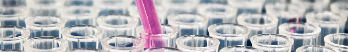
Aydogan Ozcan, Ph.D.
AIMBE College of Fellows Class of 2017 For his pioneering contributions to bio-photonics, computational imaging, sensing and diagnostics technologies impacting telemedicine, mobile-health and global health applications.
UCLA’s Virtual Histology Could Eliminate Need for Invasive Biopsies for Some Skin Conditions and Cancers
Via Dark Daily | January 19, 2022What effect would elimination of tissue biopsies have on dermatopathology and clinical laboratory revenue? Quite a lot. Dermatologists alone account for a significant portion of skin biopsies sent to dermatopathologists. Thus, any new technology that can “eliminate the need for invasive skin biopsies” would greatly reduce the number of histopathological referrals and reduce revenue to those practices.
Nevertheless, one such new technology may have been created by Ozcan Research Group in a proof-of-concept study they conducted at the University of California, Los Angeles (UCLA).
Called Virtual Histology, the technology applies artificial intelligence (AI) deep learning methods to reflectance confocal microscopy (RCM) images “to rapidly perform virtual histology of in vivo, label-free RCM images of normal skin structure, basal cell carcinoma, and melanocytic nevi with pigmented melanocytes, demonstrating similar histological features to traditional histology from the same excised tissue,” the UCLA scientists wrote in their study, published in the Nature peer-reviewed journal Light: Science and Applications… Continue reading.
New imaging technology may reduce need for skin biopsies
Via Medical Xpress | November 18, 2021Instead of surgically removing a sample of skin, sending it to a lab and waiting several days for results, your dermatologist takes pictures of a suspicious-looking lesion and quickly produces a detailed, microscopic image of the skin.
This could become routine in clinics, the result of a new “virtual histology” technology being developed by researchers at the UCLA Samueli School of Engineering and the David Geffen School of Medicine at UCLA, according to today’s article in Light: Science & Applications, a journal of the Springer Nature Group. Histology is the study of the microscopic structure of tissues.
“This process bypasses several standard steps typically used for diagnosis—including skin biopsy, tissue fixation, processing, sectioning and histochemical staining. Images appear like biopsied, histochemically stained skin sections imaged on microscope slides,” said the study’s senior author, Aydogan Ozcan… Continue reading.
Recurrent neural network advances 3D fluorescence imaging
Via EurekAlert | March 23, 2021Rapid 3D microscopic imaging of fluorescent samples has gained increasing importance in numerous applications in physical and biomedical sciences. Given the limited axial range that a single 2D image can provide, 3D fluorescence imaging often requires time-consuming mechanical scanning of samples using a dense sampling grid. In addition to being slow and tedious, this approach also introduces additional light exposure on the sample, which might be toxic and cause unwanted damage, such as photo-bleaching.
By devising a new recurrent neural network, UCLA researchers have demonstrated a deep learning-enabled volumetric microscopy framework for 3D imaging of fluorescent samples. This new method only requires a few 2D images of the sample to be acquired for reconstructing its 3D image, providing ~30-fold reduction in the number of scans required to image a fluorescent volume… Continue reading.
Deep learning enables early detection and classification of live bacteria using holography
Via Biophotonics World | July 10, 2020Waterborne diseases affect more than 2 billion people worldwide, causing substantial economic burden. For example, the treatment of waterborne diseases costs more than $2 billion annually in the United States alone, with 90 million cases recorded per year. Among waterborne pathogen-related problems, one of the most common public health concerns is the presence of total coliform bacteria and Escherichia coli (E. coli) in drinking water, which indicates fecal contamination. Traditional culture-based bacteria detection methods often take 24-48 hours, followed by visual inspection and colony counting by an expert, according to the United States Environmental Protection Agency (EPA) guidelines. Alternatively, molecular detection methods based on, for example, the amplification of nucleic acids, can reduce the detection time to a few hours, but they generally lack the sensitivity for detecting bacteria at very low concentrations, and are not capable of differentiating between live and dead microorganisms. Furthermore, there is no EPA-approved nucleic acid-based method for detecting coliform bacteria in water samples… Continue reading.
Aydogan Ozcan, Ph.D. To be Inducted into Medical and Biological Engineering Elite
Via AIMBE | March 1, 2017WASHINGTON, D.C.— The American Institute for Medical and Biological Engineering (AIMBE) has announced the pending induction of Aydogan Ozcan, Ph.D., Professor, Electrical Engineering and Bioengineering Departments, California NanoSystems Institute, University of California, Los Angeles, to its College of Fellows. Dr. Ozcan was nominated, reviewed, and elected by peers and members of the College of Fellows For his pioneering contributions to bio-photonics, computational imaging, sensing and diagnostics technologies impacting telemedicine, mobile-health and global health applications..